7th April 2025
09:20-10:00
Dr. Roeland Boer
XALOC Beamline,
ALBA Synchrotron, Spain
10:00-10:40
Dr. Lin Huang
Group Leader Guangdong-Hong Kong Joint Laboratory for RNA Medicine, Medical Research Center, Sun Yat-Sen Memorial Hospital, Sun Yat-Sen University, China
10:40-11:20
Dr.Ana Joaquina Pérez-Berna
MISTRAL Beamline Scientist,
ALBA Synchrotron, Spain
Time: 09:20-10:00_Dr. Roeland Boer
Time: 10:00-10:40_Dr. Lin Huang
Enhancing RNA Crystallization and Resolution via Some general strategies
- X-ray crystallography is a fundamental technique for determining RNA structures at atomic resolution. However, obtaining high-resolution RNA crystals remains a significant challenge. Here, we present a novel strategy to improve the resolution of RNA crystals by selectively substituting Watson–Crick base pairs with GU pairs within RNA sequences. Our approach has successfully yielded high-resolution structures for eight unique RNA crystals. Notably, six cases demonstrated marked resolution enhancement upon GC/AU to GU base pair substitution, with two instances achieving high-resolution structures from initially poor data. Interestingly, reverting GU to GC base pairs also improved resolution in one case. This method facilitated the first structural determinations of the LINE-1 and OR4K15 ribozymes, the 2′-deoxyguanosine-III riboswitch, and the Broccoli RNA aptamer. We show that placing GU base pairs within the first 5′ helical stem or in one peripheral stem of any given RNA species is sufficient. These findings provide a simple and effective approach for designing or selecting RNA sequences to facilitate high-resolution structure determination.
- Furthermore, I will also present additional methods developed in my laboratory that aid in RNA crystallization.
Time: 10:40-11:20_Dr.Ana Joaquina Pérez-Berna
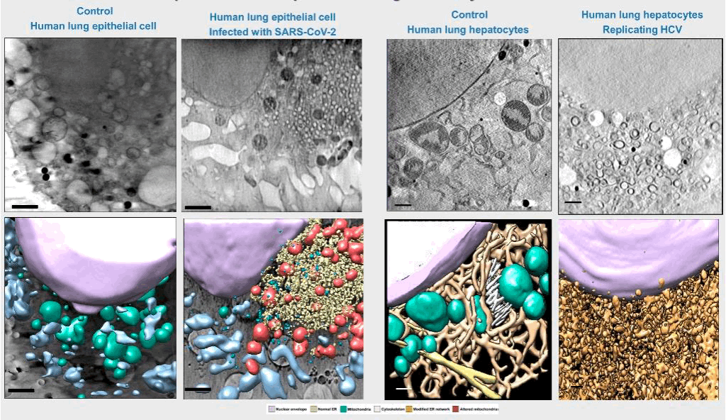
Correlative imaging of biological cells by cryo-soft-Xray-tomography
- Cryo soft X-ray tomography (SXT) of whole hydrated cells in the water window energy range can provide relevant structural information of complex cellular phenomena with chemical sensitivity at spatial resolutions of 30 nm half pitch [1]. Functional studies can be achieved by correlating this information with cryo visible light fluorescence microscopy in 2D or 3D [2, 3], as well as with 3D cryo hard X-ray fluorescence to localize and quantify specific molecules. The chemical insight into cellular aspects could be also correlated the cellular 3D structural maps with infrared microscopy, using synchrotron-based infrared microscopy (MIRAS-beamline) [4]. In addition, spectroscopic imaging can also be used to understand biomineralization processes [5]. Examples of 3D correlative cryo X-ray imaging research will be presented.

REFERENCES
[1] Schneider G. et al. Nat. Methods 7: 985-987 (2010)
[2] Pérez-Berná A.J. et al. ACS Nano 10, 6597-6611 (2016)
[3] Pérez-Berná A.J. et al. ACS Nano ACSnano, 17(22), pp. 22708–22721 (2023)
[4] Pérez-Berná A.J. et al. Acta Crystallographica Section D. ActaCryst.(2021). Nov. 2111/58(12)10
[5] Kahil K et al. PNAS 117 (49), 30957-30965 (2020).
- Our results also constitute a proof of concept for the use of cryo-SXT as a platform that enables determining the potential impact of candidate compounds on the ultrastructure of the cell that may assist drug development at a preclinical level.
Teams link for the event
7th April 2025